Tuesday, July 2, 2013

Recently banned drugs in India

The Indian government has banned three popular medicines the widely prescribed anti-diabetes drug pioglitazone, painkiller analgin and anti-depressant deanxit—in the wake of health risks associated with them.
While it's believed that pioglitazone can cause heart failure and increases the risk of bladder cancer, analgin has been discarded the world over on grounds of patient safety. Deanxit, on the other hand is a harmful combination, which has been long banned even in Denmark, its country of origin.The ministry of health and family welfare has suspended the manufacture and sale of all three drugs under Section 26A of the Drugs and Cosmetics Act, 1940 with immediate effect, through a notification issued on June 18, 2013.


During 2010-2011 , the Drug Controller General banned several drugs such as anti-diabetes medicine Rosiglitazone, anti-obesity drug Sibutramine, pain-killer Nimesulide for pediatric use, antibiotic Gaifloxacin, as well as Cisapride and Tegaserod, which are used for treating gastro-intestinal motility and irritable bowel syndrome , respectively. 
While the ban on Analgin in India has come after almost 36 years after the drug was banned in the US (which banned it in 1977), Pioglitazone was pulled out of France in 2011 for an increased risk of bladder cancer. 
 Analgin was withdrawn from Sweden in 1997 for the risk of causing a sharp fall in white blood cells, a potentially fatal condition. It is still being marketed in India, the house panel noted. The drug is also banned in France, Canada, Australia, New Zealand, Japan among a host of other countries. Pioglitazone, howevercontinues to be sold in most other major markets, including the US, the UK, Japan, Canada. 

General advise: Do not self medicate with NSAIDs* (e.g. Diclofenac, Lornoxicam, Etoricoxib etc.) Frequent use of these drugs is associated with the development of kidney and liver toxicity and causing gastric ulceration and bleeding. Hence, do not take these medications for longer than advised by your doctor.

*NSAIDs: Non-steroidal anti-inflammatory drug.

#A related blog post link: http://sciencedoing.blogspot.in/2013/03/pollution-threat-vanishing-vultures.html

#Following sites to be referred for more banned drugs in India:
 http://www.drugscontrol.org/ban_drugs.htm
 http://www.pharmatutor.org/pharmapedia/banned-drugs-in-india-manufacture-sale
 http://cdsco.nic.in/html/drugsbanned.html
 http://addictionsupport.aarogya.com/resources/733-drugs-banned-in-india.html

#News thankfully shared from:
 http://timesofindia.indiatimes.com
 http://economictimes.indiatimes.com


Tuesday, June 25, 2013

Urinary Bladder capacity, Acute and Chronic Urinary Retention causes, Benign Prostatic Hyperplasia factors and Alpha-1 blocker treatment

For the urine to exit the bladder, both the autonomically controlled internal sphincter and the voluntarily controlled external sphincter must be opened. Problems with these muscles can lead to incontinence.

The urinary bladder usually holds 300-350 ml of urine. As urine accumulates, the rugae flatten and the wall of the bladder thins as it stretches, allowing the bladder to store larger amounts of urine without a significant rise in internal pressure.
bladder picture
The urinary bladder is a muscular sac in the pelvis, just above and behind the pubic bone. When empty, the bladder is about the size and shape of a pear.
The internal sphincter is a continuation of the detrusor muscle and is made of smooth muscle, therefore it is under involuntary or autonomic control. This is the primary muscle for prohibiting the release of urine.
The external sphincter muscle of urethra  in males is a secondary sphincter to control the flow of urine through the urethra. Unlike the internal sphincter muscle, the external sphincter is made of skeletal muscle, therefore it is under voluntary control of the somatic nervous system.
share courtesy: wikipedia.org
Urine is made in the kidneys, and travels down two tubes called ureters to the bladder. The bladder stores urine, allowing urination to be infrequent and voluntary. The bladder is lined by layers of muscle tissue that stretch to accommodate urine. The normal capacity of the bladder is 400 to 600 mL.
During urination, the bladder muscles contract, and two sphincters (valves) open to allow urine to flow out. Urine exits the bladder into the urethra, which carries urine out of the body. Because it passes through the penis, the urethra is longer in men (8 inches) than in women (1.5 inches).
Urinary retention: Urine does not exit the bladder normally due to obstruction or suppressed bladder muscle activity. The bladder may swell to hold more than a quart of urine.
Causes of Urinary retention: There are two general types of urinary retention: obstructive and non-obstructive. If there is an obstruction (for example, kidney stones), urine cannot flow freely through the urinary track. Non-obstructive causes include a weak bladder muscle and nerve problems that interfere with signals between the brain and the bladder. If the nerves aren’t working properly, the brain may not get the message that the bladder is full.
Some of the most common causes of non-obstructive urinary retention are:
  • Stroke
  • Vaginal childbirth
  • Pelvic injury or trauma
  • Impaired muscle or nerve function due to medication or anesthesia
  • Accidents that injure the brain or spinal cord
Obstructive retention may result from:
  • Cancer
  • Kidney or bladder stones
  • Enlarged prostate (BPH) in men    
Acute and Chronic urine retention: Urine retention may be acute or chronic and occurs when something prevents the flow of urine from the bladder to the urethra and out of the body. 

In acute urine retention, but the person is unable to pass the urine, resulting in significant pain. 
Chronic urine retention is where a person is able to pass small quantities of urine with difficulty and the bladder never completely empties. It is usually painless. Symptoms of acute urine retention include lower abdominal pain, a sensation of fullness in the lower abdomen, and a painful urge and inability to pass urine. Chronic urine retention symptoms include swelling of the abdomen, a frequent need to urinate, difficulty in starting the urine flow, a weak urine flow, and dribbling at the end of urinating and between urinating. Treatment options include temporary or permanent catheterization, medication, treating the underlying cause, herbal therapies, and homoeopathy.

Acute urinary retention (AUR) is the sudden inability to pass urine. It is usually painful and requires emergency treatment with a urinary catheter. 
Causes: These are numerous and can be classified as:
  • In men - benign prostatic hypertrophy (BPH), meatal stenosis, paraphimosis, penile constricting bands, phimosis, prostate cancer.
  • In women - prolapse (cystocele, rectocele, uterine), pelvic mass (gynaecological malignancy, uterine fibroid, ovarian cyst), retroverted gravid uterus.
  • In both - bladder calculi, bladder cancer, faecal impaction, gastrointestinal or retroperitoneal malignancy, urethral strictures, foreign bodies, stones.
Infectious and inflammatory:
  • In men - balanitis, prostatitis and prostatic abscess.
  • In women - acute vulvovaginitis, vaginal lichen planus and lichen sclerosis, vaginal pemphigus.
  • In both - bilharzia, cystitis, herpes simplex virus (particularly primary infection), peri-urethral abscess, varicella-zoster virus.
A healthy human prostate is classically said to be slightly larger than a walnut. The mean weight of the "normal" prostate in adult males is about 11 grams, usually ranging between 7 and 16 grams. It surrounds the urethra just below the urinary bladder and can be felt during a rectal exam. It is the only exocrine organ located in the midline in humans and similar animals. share courtesy: wikipedia.org


#BPH is by far the most common cause of urinary retention. Alpha-1 blockers given before catheter removal increase the chances of a successful TWOC.
Diagram illustrating normal prostate (left)
and benign prostatic hyperplasia (right).
 share courtesy: http://en.wikipedia.org
Benign Prostatic Hyperplasia: BPH involves hyperplasia of prostatic stromal and epithelial cells, resulting in the formation of large, fairly discrete nodules in the periurethral region of the prostate. When sufficiently large, the nodules compress the urethral canal to cause partial, or sometimes virtually complete, obstruction of the urethra, which interferes with the normal flow of urine. It leads to symptoms of urinary hesitancy, frequent urination, dysuria (painful urination), increased risk of urinary tract infections, and urinary retention. Although prostate specific antigen levels may be elevated in these patients because of increased organ volume and inflammation due to urinary tract infections, BPH does not lead to cancer or increase the risk of cancer.
BPH involves hyperplasia (an increase in the number of cells) rather than hypertrophy (a growth in the size of individual cells), but the two terms are often used interchangeably, even amongst urologists. 
Its prevalence is age dependent. Histological evidence of BPH is rarely observed in men under 50 years of age, but by age 80 virtually all men will have some histological evidence of the process. It is unclear what specific factors regulate the degree of hyperplasia, which ultimately dictates the size of the prostate gland, nor is there any consensus regarding the prostate size that qualifies for the diagnosis of benign prostatic enlargement (BPE).


Alpha blockers for the treatment of Benign Prostatic Hyperplasia:
The relative degree of stromal and epithelial hyperplasia is highly variable. Overall, approximately 80% and 20% of the hyperplastic volume is composed of stromal and epithelial elements, respectively. Half of the stromal hyperplasia is composed of smooth-muscle elements. For decades, it was assumed that the enlarged hyperplastic prostate caused BOO via both dynamic and static mechanisms. The dynamic obstruction was thought to be the result of smooth-muscle hyperplasia causing a functional obstruction and static obstruction arising from the bulk enlargement of the hyperplastic process encroaching upon the prostatic urethra.

Marco Caine demonstrated in 1975 that strips of human prostate contracted in response to norepinephrine.The norepinephrine-induced contractions were inhibited by pretreatment with phenoxybenzamine, a non-selective inhibitor of alpha adrenoceptor. These studies implicated the alpha adrenoceptor as the mediator of prostate smooth-muscle contraction. Lepor and Shapiro were the first investigators to characterize both alpha 1 and 2 adrenoceptors in the human prostate using radioligand binding studies.  

Tamsulosin was the third alpha 1 blocker to be approved for the treatment of BPH. Tamsulosin was brought to market as the first subtype selective alpha 1 antagonist for the treatment of BPH. Tamsulosin alpha 1 subtype selective was supported by binding studies, which showed that tamsulosin was approximately tenfold more selective for the alpha 1a versus alpha 1b subtype. There was no demonstrable subtype selectivity of tamsulosin for the alpha 1a versus alpha 1d subtypes. The modest receptor selectivity of tamsulosin is not sufficient to result in a clinically meaningful advantage. Typically, clinical advantages attributed to pharmacological selectivity require a receptor selectivity well beyond the tenfold difference observed with tamsulosin.
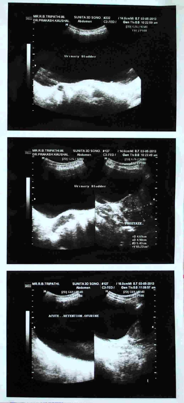
A case report:
Age: 86 years male,
Acute urinary retention 1400 ml,
Prostate enlarge, 65 gm in weight,
Chronic retention of urine
cause may be BPH with
Bladder Neck Obstruction.
#As seen in above sonograph 
picture
Two pivotal trials of tamsulosin supported the NDA for the treatment of symptomatic BPH. Both 0.4 and 0.8 mg of tamsulosin achieved clinically significant improvements in symptom scores and peak flow rate. The ability of the 0.4 mg tamsulosin dose to achieve a clinically significant effect without the requirement for dose titration represented a unique advantage over the other approved alpha blockers. Although a 0.8 mg daily dose was more effective than 0.4 mg, it did not gain popularity because it required both dose titration and taking 2 tablets of 0.4 mg. (A 0.8 mg tablet was not commercially available.) The primary reason tamsulosin was prescribed over terazosin and doxazosin was not due to greater efficacy or better tolerability, but simply the lack of dose titration. The prescribing community placed a greater value on eliminating the dose response at the expense of increasing the incidence of ejaculatory dysfunction, which was thought to be retrograde ejaculation as a result of relaxation of the bladder neck. Recent studies have demonstrated that tamsulosin causes anejaculation rather than retrograde ejaculation. The mechanism for the increased incidence of ejaculatory dysfunction associated with tamsulosin has been attributed to its affinity for dopaminergic and other central nervous system receptors. 
An episode of acute urinary retention is no longer an absolute indication for surgical intervention. Alpha blockers are a very reasonable initial option for managing acute urinary retention.

Following sites referred thankfully and reference for further detail:
http://en.wikipedia.org
http://www.webmd.com
http://www.medtronic.com
http://www.naturaltherapypages.com.au/glossary/Urine_Retention#ixzz2T2pDuWCp 
http://www.patient.co.uk/doctor/acute-urinary-retention
NCBI journal Rev Urol. 2007 Fall; 9(4): 181–190
file:///E:/Alpha%20Blockers%20for%20the%20Treatment%20of%20Benign%20Prostatic%20Hyperplasia.htm


Some abbreviations used:
Food and Drug Administration (FDA)
lower urinary tract symptoms (LUTS)
bladder outlet obstruction (BOO)
benign prostatic enlargement (BPE)
benign prostatic hyperplasia (BPH)
Medical Therapy of Prostatatic Symptoms (MTOPS)
The American Urological Association Symptoms Index (AUASI)
International Prostate Symptom Index Score (IPSS)
new drug application (NDA)
Three subtypes of the alpha 1 adrenoceptor (alpha 1a, alpha 1b, alpha 1d) have been cloned and  pharmacologically characterized.
The Antihypertensive and Lipid-Lowering Treatment to Prevent Heart Attack Trial (ALLHAT)

Tuesday, June 18, 2013

Drinking too much water can lead to hyponatremia, water intoxication, water poisoning and death

Liquid H2O is the sine qua non of life.  
Making up about 66 percent of the human body, water runs through the blood, inhabits the cells, and lurks in the spaces between. At every moment water escapes the body through sweat, urination, defecation or exhaled breath, among other routes. Replacing these lost stores is essential but rehydration can be overdone. There is such a thing as a fatal water overdose.  

The poison is the dose
Water can cause the brain to swell 
share courtesy: http://news.bbc.co.uk

Drinking several litres over a relatively short period of time could be enough to cause water intoxication. Those most at risk include people taking ecstasy, as the drug increases thirst and facilitates the release of anti-diuretic hormones so more water is taken in but cannot be excreted. Also, elderly people because their kidney function may be impaired. 

Strange but true; drinking too much water can kill.
In a hydration-obsessed culture, people can and do drink themselves to death. 


As long as you are healthy and equipped with a thirst barometer unimpaired by old age or mind-altering drugs, follow Verbalis's advice, "drink to your thirst. It's the best indicator." 

Beware of mindlessly drinking several glasses of water per day without considering your diet, exercise habits, climate, and sense of thirst. And when you do find yourself in need of water, remember that you can get it from liquids and/or whole foods that are rich in water.

Forcing your body to accept a large amount of water within a short period of time - say, an hour or two - can be fatally dangerous to your health. Here's why:
If you force large amounts of water into your system over a short period of time, your kidneys will struggle to eliminate enough water from your system to keep the overall amount at a safe level.
As your circulatory system becomes diluted with excess water, the concentration of electrolytes in your blood will drop relative to the concentration of electrolytes in your cells. In an effort to maintain an equal balance of electrolytes between your blood and your cells, water will seep into your cells from your blood, causing your cells to swell.
If this swelling occurs in your brain, you'll experience increased intracranial pressure i.e. your brain will get squeezed because the flat bones that make up your skull don't provide much give. Depending on how much water your drink in a short period of time, you could experience a wide variety of symptoms, ranging from a mild headache to impaired breathing. As occurred in the tragic water-drinking contest, it's quite possible to die if you drink enough water in a short period of time. 
Water intoxication, also known as water poisoning or dilutional hyponatremia, is a potentially fatal disturbance in brain functions that results when the normal balance of electrolytes in the body is pushed outside safe limits by over-hydration.

Water, just like any other substance, can be considered a poison when over-consumed in a specific period of time. Water intoxication mostly occurs when water is being consumed in a high quantity without giving the body the proper nutrients it needs to be healthy.

One of the many Risk factors, is  Psychogenic polydipsia is the psychiatric condition in which patients feel compelled to drink large quantities of water, thus putting them at risk of water intoxication. This condition can be especially dangerous if the patient also exhibits other psychiatric indications (as is often the case), as the care-takers might misinterpret the hyponatremic symptoms.

Following sites referred thankfully and reference for further detail:
wikipedia.org
http://www.scientificamerican.com
http://news.bbc.co.uk
http://drbenkim.com/drink-too-much-water-dangerous.html

Tuesday, June 11, 2013

Human brain: Hypothalamus as control center for ageing identified but risk lies in follow up

Hypothalamus region of the brain is the control centre of ageing. Dongsheng Cai from the Albert Einstein College of Medicine in New York has discovered yet another vital duty on the hypothalamus’ already impressive CV—it’s a command centre that coordinates the ageing process across the entire body. By manipulating proteins within it, Cai managed to speed up or slow down the pace of ageing in mice. 
The brain's mechanism for controlling ageing
has been discovered – and manipulated to
shorten and extend the lives of mice.
Drugs to slow ageing could follow
share courtesy:
http://www.newscientist.com/section/science-news
Cai’s team found that the ageing hypothalamus builds up more microglia—a dedicated brand of inflammatory immune cells in the brain, which destroy infectious microbes and vacuum up debris. They also produce NF-kB, which tells neighbouring neurons to do the same. This flood of NF-kB switches off the gene for a hormone called GnRH.
That’s a surprise—GnRH is best known as a reproductive hormone. It controls the development of our sexual organs and the making of eggs and sperm. 

Human hypothalamus 
(animation, shown in red)
share courtesy: wikipedia.org
Howard Chang from Stanford University says, “If reproduced by others, this work provides further evidence that aging is a regulated rather than a [random] process.” 

But let’s recap what we already know, just to appreciate how bizarre this chain of results is. You have a life-support centre in the brain, a protein that coordinates the immune system, and a hormone that controls our sex organs, all working together to influence… the pace of ageing?

This research follow up may bring some serious consequences as: 
Given the pathway, we would not be surprised if a large experiment showed a significant increase of cancer risk. NF-kB is a well known cancer pathway, lower levels of NF-kB1 allow cancer cells to avoid cell death (apoptosis). I imagine this works along the same lines.  

Following sites referred thankfully and reference for further detail:
http://phenomena.nationalgeographic.com/2013/05/01/almond-sized-brain-region-is-control-centre-for-ageing/
mb, may1, 2013 on http://phenomena.nationalgeographic.com/2013/05/01/almond-sized-brain-region-is-control-centre-for-ageing/
http://www.newscientist.com/section/science-news
wikipedia.org

Tuesday, June 4, 2013

Images of a molecule

"Nobody has ever taken direct, single-bond-resolved images of individual molecules, right before and immediately after a complex organic reaction," says Felix Fischer of the U.S. Department of Energy's Lawrence Berkeley National Laboratory. 
(The researchers report their results in the June 7, 2013 edition of the journal Science, available in advance on Science Express.)

Almost as clearly as textbook diagram, this image made by a noncontact atomic force microscope reveals the positions of individual atoms and bonds, in a molecule having 26 carbon atoms and 14 ydrogen atoms structured as three connected benzene rings. Credit: Lawrence Berkeley national Laboratory and University of California at Berkeley

The original reactant molecule, resting on a flat silver surface, is imaged both before and after the reaction, which occurs when the temperature exceeds 90 degrees Celcius. The two most common final products of the reaction are shown. The three-angstrom scale bars (an angstrom is a ten-billionth of a meter) indicate that the reactant and product molecules are about a billionth of a meter across. Credit: Lawrence Berkeley national Laboratory and University of California at Berkeley

The single-atom tip of the noncontact atomic force microscope "feels" changes in the strength of electronic forces as it moves across the surface at a constant height. Resulting movements of the stylus are detected by a laser beam to compute images. Credit: Lawrence Berkeley national Laboratory and University of California at Berkeley
#thankfully shared from: http://phys.org
May 30, 2013


Tuesday, May 28, 2013

Photosynthesis: new insights

Photosynthesis: Lessons from nature

Photosynthesis is one of nature's finest miracles. Through the photosynthetic process, green plants absorb sunlight in their leaves and convert the photonic energy into chemical energy that is stored as sugars in the plants' biomass. If we can learn from nature and develop an artificial version of photosynthesis we would have an energy source that is absolutely clean and virtually inexhaustible. (September 23, 2011)

Through the miracle of photosynthesis, plants absorb sunlight in their leaves and convert the photonic energy into chemical energy that is stored as sugars in the plants' biomass. (Credit: Photo by Roy Kaltschmidt, Berkeley Lab)

"Solar energy is forecasted to provide a significant fraction of the world's energy needs over the next century, as sunlight is the most abundant source of energy we have at our disposal," says Graham Fleming, Vice Chancellor for Research at the University of California (UC) Berkeley who holds a joint appointment with Lawrence Berkeley National Laboratory (Berkeley Lab). 

"However, to utilize solar energy harvested from sunlight efficiently we must understand and improve both the effective capture of photons and the transfer of electronic excitation energy." 


Photosynthesis: Evolutionary perspective

Photosynthesis is one of the fundamental processes of life on Earth. The evolutionary transition from anoxygenic (no oxygen produced) to oxygenic (oxygen-producing) photosynthesis resulted in the critical development of atmospheric oxygen in amounts large enough to allow the evolution of organisms that use oxygen, including plants and mammals.(April 3, 2012)

Ferns. Photosynthesis is one of the fundamental processes of life on Earth. The evolutionary transition from anoxygenic (no oxygen produced) to oxygenic (oxygen-producing) photosynthesis resulted in the critical development of atmospheric oxygen. One of the outstanding questions of the early Earth is how ancient organisms made this transition. (Credit: © Pontus Edenberg / Fotolia)

Plants and algae, as well as cyanobacteria, use photosynthesis to produce oxygen and "fuels," the latter being oxidizable substances like carbohydrates and hydrogen. There are two pigment-protein complexes that orchestrate the primary reactions of light in oxygenic photosynthesis: photosystem I and photosystem II.

"In photosynthesis, the oxygen is produced at a special metal site containing four manganese and one calcium atom connected together as a metal cluster," explains professor James Allen. "This cluster is bound to the protein called photosystem II that provides a carefully controlled environment for the cluster."

On illumination, two water molecules bound at the cluster are split into molecular oxygen and four protons. Since water molecules are very stable, this process requires that the metal cluster be capable of efficiently performing very energetic reactions.
Allen, Williams and coworkers are trying to understand how a primitive anoxygenic organism that was capable of performing only simple low energy reactions could have evolved into oxygen-producing photosynthesis.
They have been manipulating the reaction center of the purple bacterium Rhodobacter sphaeroides encouraging it to acquire the functions of photosystem II. In the recent publication, they describe how a mononuclear manganese bound to the reaction center has gained some of the functional features of the metal cluster of photosystem II.
Although the mononuclear manganese cannot split water, it can react with reactive oxygen species to produce molecular oxygen. These results suggest that the evolution of photosynthesis might well have proceeded through intermediates that were capable of oxygen production and served until a protein with a bound manganese-calcium cluster evolved. 


Carotenoids Can Capture Blue/Green Light and Pass Energy On to Chlorophylls 

Pigments found in plants and purple bacteria employed to provide protection from sun damage do more than just that. Researchers from the University of Toronto and University of Glasgow have found that they also help to harvest light energy during photosynthesis. (April 4, 2013) 

Advanced optical probes using femtosecond lasers enable light harvesting processes to be examined in exquisite detail. Anticlockwise from top right: Purple bacteria and the structure of the light harvesting complex that gives these cells their distinctive purple color. This special protein incorporates molecules of bacteriochlorophyll and carotenoid to capture the energy from sunlight. The lower part of the figure shows the protein data recorded from two-dimensional laser spectroscopy. (Credit: Evgeny Ostroumov)

A series of experiments showed that a special "dark state" of the carotenoid -- a hidden level not used for light absorption at all -- acts as a mediator to help pass the energy it absorbs very efficiently to a chlorophyll pigment.

Says Scholes. "It is amazing that nature uses so many aspects of a whole range of quantum mechanical states in carotenoid molecules, moreover, and puts those states to use in such diverse ways."

  cientists Swap Key Metal Necessary For Turning Sunlight Into Chemical Energy

Scientists Swap Key Metal Necessary For Turning Sunlight Into Chemical Energy

Photosynthesis is a remarkable biological process that supports life on earth. Plants and photosynthetic microbes do so by harvesting light to produce their food, and in the process, also provide vital oxygen for animals and people.(May 23, 2009)

The reactions that convert light to chemical energy happen in a millionth of a millionth of a second, which makes experimental observation extremely challenging. A premier ultrafast laser spectroscopic detection system established at the Biodesign Institute, with the sponsorship of the National Science Foundation, acts like a high-speed motion picture camera. It splits the light spectrum into infinitesimally discrete slivers, allowing the group to capture vast numbers of ultrafast frames from the components of these exceedingly rapid reactions. These frames are then mathematically assembled, allowing the group to make a figurative "movie" of the energy transfer events of photosynthesis. (Credit: Arizona State University Biodesign Institute)

In all plant chlorophylls, only one particular metal, magnesium, is held tightly within the molecule's center.

During photosynthesis, plants have two photosystems that work in tandem: photosystem I and photosystem II. To peer at the inner workings of photosynthesis, the team used a hardy, well-studied, photosynthetic bacterium called Rhodobacter sphaeroides. An organism similar to this purple bacterium was likely one of the earliest photosynthetic bacteria to evolve. The purple bacteria possess a simplified system similar to photosystem II.

The center stage of photosynthesis is the reaction center, where light energy is funneled into specialized chlorophyll binding proteins. The research team had previously demonstrated that the movement of the reaction center proteins during photosynthesis facilitates the light-driven movement of electrons between molecules in the reaction center, helping the plant or bacteria to harness light energy efficiently even if conditions aren't optimal. Every time the team introduced disruptions into this electron pathway, the proteins were able to compensate by moving and energetically guiding the electrons through their biological circuit. 


Photosynthesis: A new source of electrical energy
Biofuel cell works in cactus

Scientists in France have transformed the chemical energy generated by photosynthesis into electrical energy by developing a novel biofuel cell. The advance offers a new strategy to convert solar energy into electrical energy in an environmentally-friendly and renewable manner. In addition, the biofuel cell could have important medical applications. (February 18,2008) 

Biofuel cell inserted in a cactus and graph showing the course of electrical current as a function of illumination of the cactus (black: glucose, red: O2).

The researchers showed that a biofuel cell inserted in a cactus leaf could generate power of 9 μW per cm2. Because this yield was proportional to light intensity, stronger illumination accelerated the production of glucose and O2 (photosynthesis), so more fuel was available to operate the cell. In the future, this system could ultimately form the basis for a new strategy for the environmentally-friendly and renewable transformation of solar energy into electrical energy. 


Artificial Photosynthesis 

Researchers from the Department of Chemistry at the Royal Institute of Technology (KTH) in Stockholm, Sweden, have managed to construct a molecular catalyzer that can oxidize water to oxygen very rapidly. In fact, these KTH scientists are the first to reach speeds approximating those is nature's own photosynthesis. The research findings play a critical role for the future use of solar energy and other renewable energy sources. (April 12, 2012)

Grass. Scientists have imitated natural photosynthesis and created a record-fast molecular catalyzer. (Credit: © Nejron Photo / Fotolia)

Researchers all over the world, including the US, Japan, and the EU, have been working for more than 30 years on refining an artificial form of photosynthesis. The results have varied, but researchers had not yet succeeded in creating a sufficiently rapid solar-driven catalyzer for oxidizing water.

But now, together with research colleagues, he has imitated natural photosynthesis and created a record-fast molecular catalyzer. The speed with which natural photosynthesis occurs is about 100 to 400 turnovers per seconds. The KTH have now reached over 300 turnovers per seconds with their artificial photosynthesis.

"This is clearly a world record, and a breakthrough regarding a molecular catalyzer in artificial photosynthesis," says Licheng Sun.

"When it comes to renewable energy sources, using the sun is one of the best ways to go," says Sun. 


Artificial Leaf

Scientists have claimed one of the milestones in the drive for sustainable energy -- development of the first practical artificial leaf. Speaking in Anaheim, California at the 241st National Meeting of the American Chemical Society, they described an advanced solar cell the size of a poker card that mimics the process, called photosynthesis, that green plants use to convert sunlight and water into energy. (March 28, 2011)

Living tree leaves. Scientists have just claimed one of the milestones in the drive for sustainable energy -- development of the first practical artificial leaf. (Credit: iStockphoto)


"A practical artificial leaf has been one of the Holy Grails of science for decades," said Daniel Nocera, Ph.D., who led the research team. "We believe we have done it. The artificial leaf shows particular promise as an inexpensive source of electricity for homes of the poor in developing countries. Our goal is to make each home its own power station," he said. "One can envision villages in India and Africa not long from now purchasing an affordable basic power system based on this technology."


Following sites referred thankfully and reference for further detail:

http://www.sciencedaily.com, the journal Analytical Chemistry.

Related blog post link:  

http://sciencedoing.blogspot.in/2013/02/chloroplast-cell-organelle-symbiotic.html

Tuesday, May 21, 2013

Oxidative stress, strain as predisposing factor in pathogenesis

Oxidative stress  
is considered to be involved in a multitude of pathogenic processes 
and is also implicated in the process of aging. 

For the first time, scientists of the  
German Cancer Research Center 
(Deutsches Krebsforschungszentrum, DKFZ) 
have been able to directly observe  
oxidative changes in a living organism. 

Their findings in fruit flies raise doubts about the validity of some widely held hypotheses: 
The research team has found no evidence that the life span is limited by the production of  
harmful oxidants.

Even though comprehensive studies have failed to provide proof until the present day,  
antioxidants are often advertised as a protection against oxidative stress and, thus, health-promoting. 
Dick and colleagues fed their flies with  
N-acetyl cysteine (NAC), 
a substance which is attributed  
an antioxidant effect 
and which some scientists consider suitable for protecting the body against presumably dangerous oxidants. 
Interestingly, no evidence of a decrease in oxidants was found in the NAC-fed flies. 
On the contrary, the researchers were surprised to find that NAC prompted the energy plants of various tissues to significantly increase oxidant production.
Yellow light signals emitted by the biosensor indicate oxidant production 
in the tissue of a migrating fly larva. 
(Credit: Tobias Dick, German Cancer 
Research Center)

 "Many things we observed in the flies with the help of the biosensors came as a surprise to us. It seems that many findings obtained in isolated cells cannot simply be transferred to the situation in a living organism," said Tobias Dick, summarizing their findings. "The example of NAC also shows that we are currently not able to predictably influence oxidative processes in a living organism by pharmacology," he adds. "Of course, we cannot simply transfer these findings from fly to man. Our next goal is to use the biosensors to observe oxidative processes in mammals, especially in inflammatory reactions and in the development of tumors."

Related blog post links for further textual matter: 
http://sciencedoing.blogspot.in/2011/12/oxygen-necessary-evil.html
Following sites referred thankfully and reference for further detail: 
#thankfully shared from http://www.sciencedaily.com

Brain waves

Brain waves   Brain cells communicate both sensory and motor impulses with the help of electrical current in all states of life i.e. resting...